Illuminating the Core of Diagnostic Imaging

Comprehensive analysis of structural modalities in medical radiography. From standard flat-panel detectors to multi-slice computed tomography, we detail the hardware and physics driving modern non-invasive diagnostics.

1895
Year of X-ray discovery by Röntgen
< 1 mSv
Average effective dose for chest DR
0.6 mm
Typical focal spot size in mammography
256+
Detector rows in modern MDCT

Primary Modalities

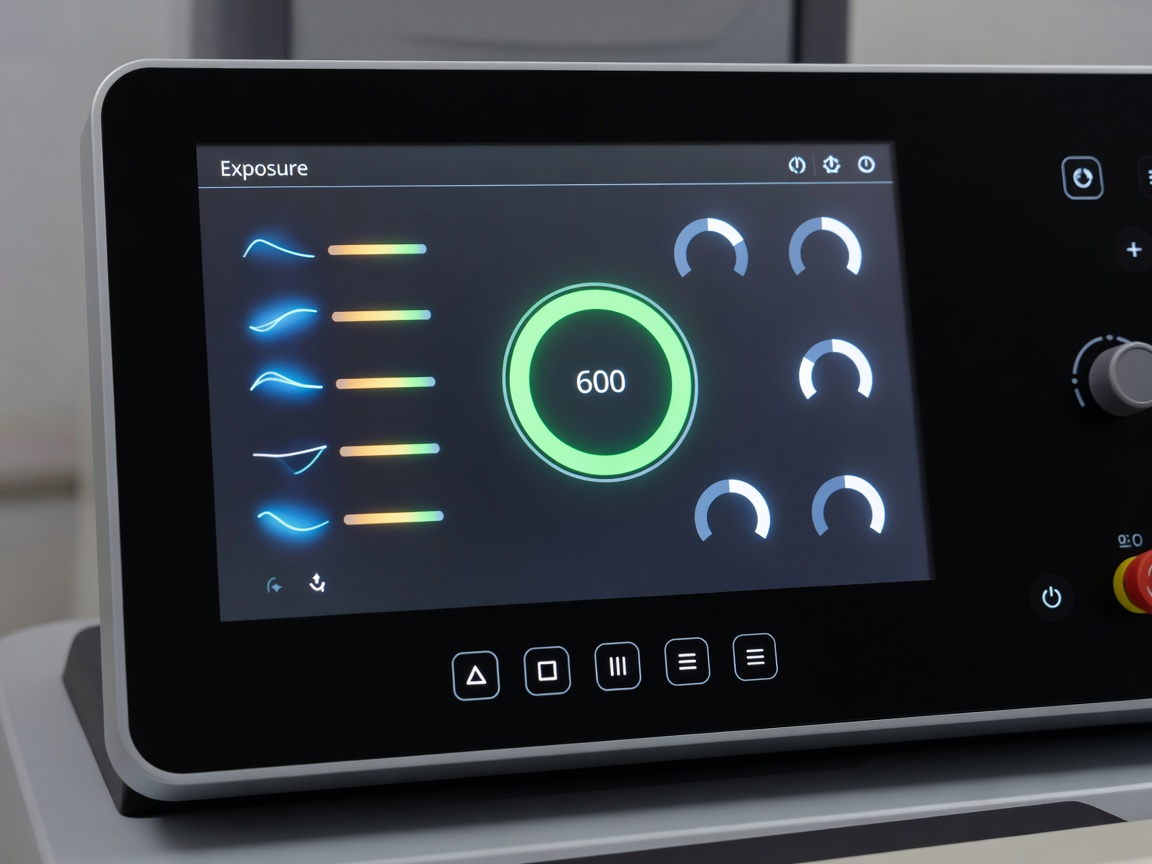
Control panel of a digital radiography system showing exposure settings
Projection Radiography General Purpose

Digital Radiography (DR)

The standard for general planar imaging. Utilizing amorphous silicon TFT arrays and cesium iodide scintillators to convert X-ray photons directly into digital signals, drastically reducing acquisition time and patient dose compared to legacy film-screen or computed radiography (CR) systems.

Explore DR Specifications
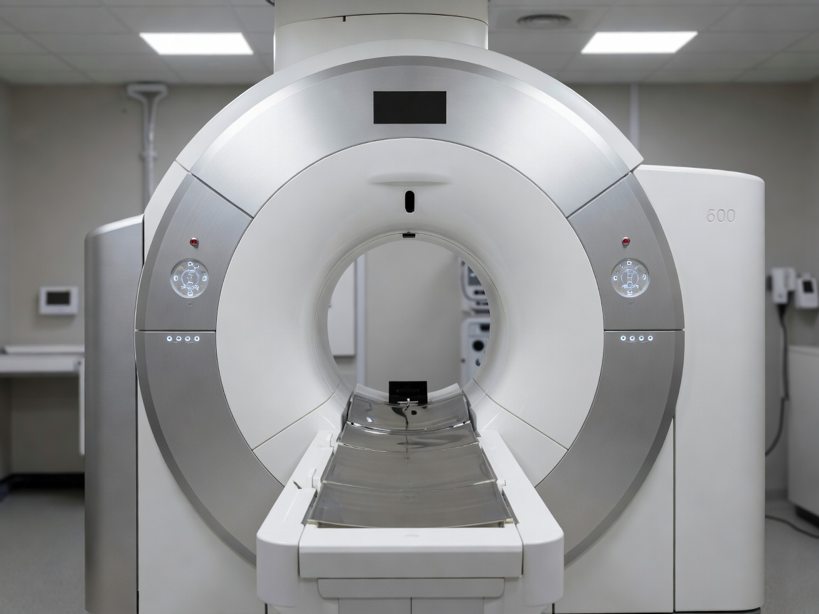
Gantry of a modern computed tomography scanner
Cross-Sectional High Contrast

Computed Tomography (CT)

Advanced volumetric imaging utilizing a continuously rotating gantry powered by slip-ring technology. Modern Multi-Detector CT (MDCT) systems acquire hundreds of overlapping slices per rotation, enabling isotropic 3D reconstruction essential for trauma, oncology, and vascular assessments.

Review CT Architecture
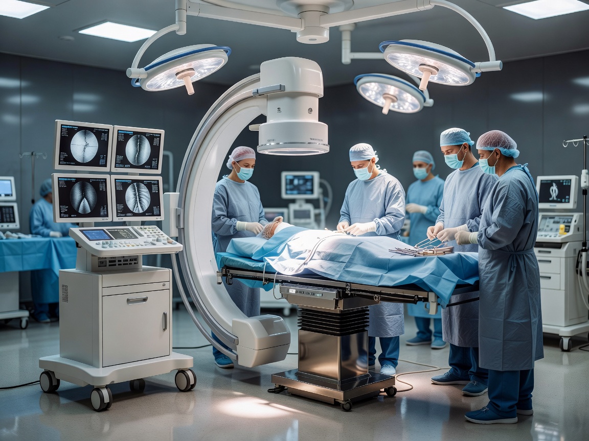
Mobile C-arm fluoroscopy unit positioned in an operating theater
Dynamic Imaging Interventional

Fluoroscopy Systems

Designed for real-time visualization of internal structures and motion. Transitioning from image intensifiers to dynamic flat-panel detectors, these continuous-beam systems are critical for angiography, barium studies, and guiding orthopedic interventions via mobile C-arms.

Analyze Fluoroscopy Tech
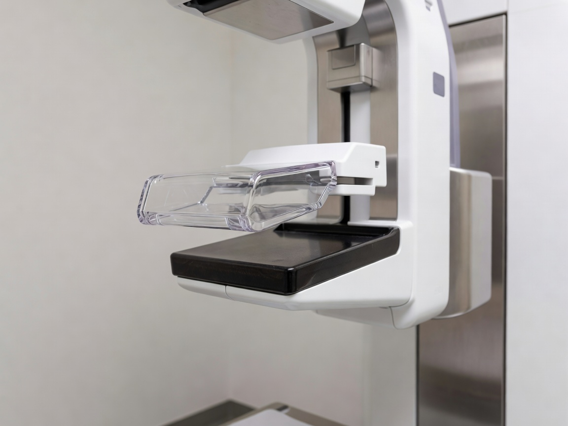
Mammography unit showing the compression paddle and detector housing
Low kVp Soft Tissue

Mammography Units

Highly specialized equipment optimized for soft tissue contrast. Operating at low peak kilovoltage (kVp) with specific target/filter combinations (like Molybdenum/Rhodium), these units maximize the detection of microcalcifications. Includes advancements in Digital Breast Tomosynthesis (DBT).

Examine Mammography Specs
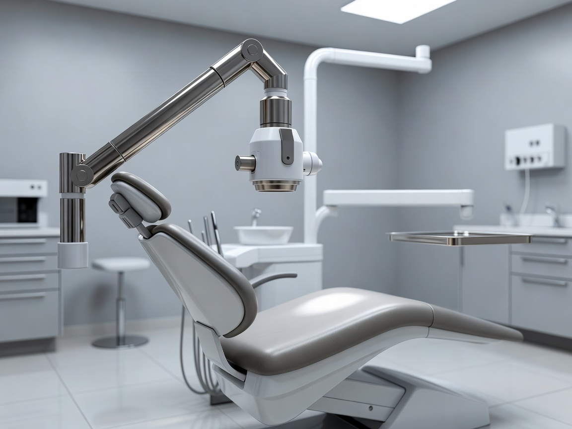
Dental intraoral X-ray tube head positioned next to a patient chair
Maxillofacial CBCT

Dental X-Ray Modalities

Ranging from compact intraoral CMOS sensors for bitewing diagnostics to extraoral panoramic and Cone Beam Computed Tomography (CBCT) systems. CBCT offers specialized 3D volumetric data crucial for implant planning and endodontics, utilizing a divergent pyramidal beam.

Discover Dental Imaging

Radiation Physics and Detector Innovation

The core principle across all X-ray modalities involves thermionic emission at a cathode, accelerating electrons across a vacuum tube to strike an anode target, generating Bremsstrahlung and characteristic X-rays. The distinction between systems lies heavily in the geometry of the beam, the filtering applied, and the detector technology.

Historically, analog film dictated exposure requirements. Today, digital solid-state detectors dominate. The shift to Indirect Conversion (using scintillators like Cesium Iodide coupled with photodiode arrays) and Direct Conversion (using Amorphous Selenium to convert X-rays directly to charge) has fundamentally altered the dose-to-image-quality ratio. This digital foundation allows for sophisticated post-processing algorithms, dual-energy subtraction, and seamless integration with PACS (Picture Archiving and Communication Systems) networks.